Neurosurgery: Understanding Common Brain And Spine Procedures
Planning and imaging frameworks for brain and spine procedures
Preoperative planning commonly begins with detailed imaging to characterize lesion size, location, and relation to surrounding structures. MRI provides soft-tissue contrast useful for tumor and cord pathology, while CT better depicts osseous detail relevant to spinal instrumentation or skull base anatomy. Vascular studies such as CTA, MRA, or catheter angiography may be necessary for vascular lesions. These data sets are often integrated into planning workstations and neuronavigation systems to visualize surgical corridors and to estimate resection margins or fixation trajectories.
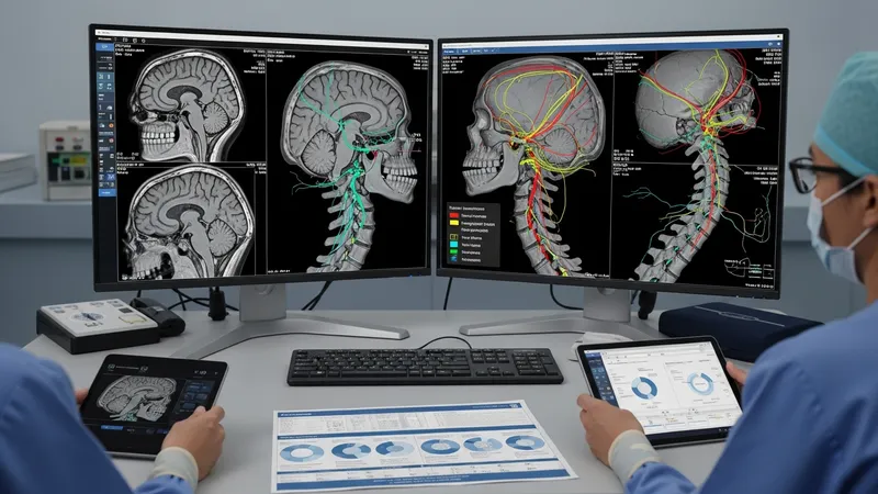
Functional imaging and mapping may be used in selected cases where preserving neurologic function is a priority. Functional MRI and tractography can illustrate likely pathways for language or motor tracts and may inform whether awake mapping or intraoperative stimulation is needed. Risk assessment also includes medical comorbidity evaluation, anticoagulation management, and considerations about prior radiation or surgery that may affect tissue planes and healing potential. Multidisciplinary planning meetings can help align goals and expectations.
Three-dimensional modeling and simulation are increasingly used for complex cranial and spinal deformity planning; these techniques may facilitate rehearsal of osteotomies or hardware placement in some centers. For endovascular planning, digital subtraction angiography provides fine vascular detail to guide catheter-based strategies. Planning frameworks typically prioritize patient safety and functional preservation, using imaging to reduce uncertainty about critical relationships and access routes before entering the operating room.
Informed consent discussions frequently reflect imaging findings and procedural alternatives, addressing potential benefits and limitations in nonabsolute terms. Documentation often includes anticipated hospital course and follow-up imaging plans to monitor for residual disease, hardware position, or vascular recurrence when applicable. Such structured planning aims to align the surgical strategy with measurable anatomic goals and the patient’s clinical priorities.