Artificial Intelligence In Breast Cancer: Applications In Diagnostic Processes
AI-Assisted Image Analysis Methods in Breast Cancer
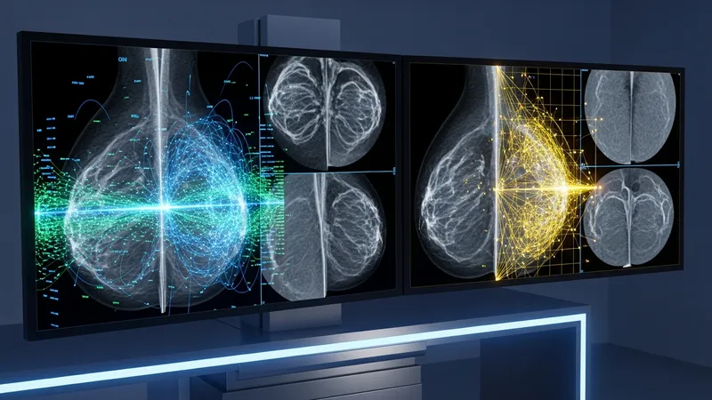
Convolutional neural networks (CNNs) are among the most widely used techniques in AI-driven breast cancer imaging. These models automate the extraction of visual features from scans, learning to recognize subtle differences in tissue composition and structure. Their architecture, characterized by multiple algorithmic layers, can process complex patterns with high granularity, which is critical for mammography and tomosynthesis evaluation.
Another prevalent method involves the use of support vector machines (SVMs) in combination with handcrafted feature extraction. Radiomics approaches often calculate statistical descriptors—such as texture, shape, and edge features—from segmented images. These features are then classified by the AI to assign probabilities or scores related to specified clinical findings. SVMs are valued for their interpretability and relatively quick training on smaller datasets.
Beyond static image review, some AI platforms facilitate temporal image analysis, comparing scans from multiple time points to detect changes over time. By analyzing progression patterns, these techniques may assist clinicians in monitoring follow-up images, especially in patients with previous abnormal findings. Such longitudinal assessment is often used as a supportive tool alongside traditional evaluation practices.
Integration with picture archiving and communication systems (PACS) is a key consideration for AI-assisted image review. Many solutions are designed to be compatible with existing clinical IT infrastructure, streamlining the delivery of AI-processed results to radiologists. This integration can help ensure that new technologies are incorporated into standard diagnostic workflows with minimal disruption.