Cardiac Surgery: Overview Of Common Procedures And Approaches
Cardiac Surgery: Surgical Planning and Preoperative Assessment
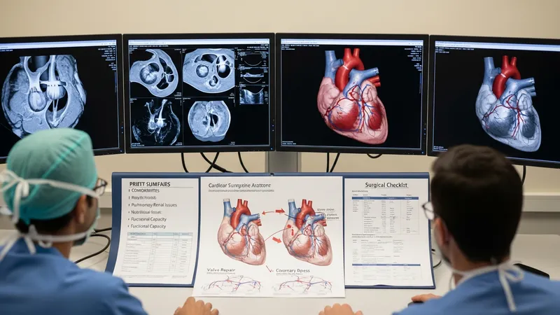
Surgical planning typically begins with detailed imaging and physiologic assessment to delineate anatomy and operative risks. Echocardiography commonly provides valve morphology and function data, while coronary angiography maps coronary arterial disease. Computed tomography or cardiac magnetic resonance imaging may be used for structural detail in complex cases. Preoperative assessment often includes evaluation of comorbidities such as pulmonary or renal disease, nutritional status, and functional capacity, all of which can influence approach selection and perioperative goals.
Multidisciplinary case review is a frequent planning element in cardiac surgical practice. Teams that include cardiac surgeons, interventional cardiologists, anaesthesiologists, and imaging specialists may discuss procedural options, potential need for adjunctive technologies, and perioperative resource planning. This collaborative approach often aims to align procedural choice with patient-specific anatomic and clinical factors, though the exact composition of teams and processes can vary between centers.
Preoperative risk mitigation may focus on optimizing reversible conditions and clarifying expectations for recovery. Examples of considerations include adjusting medications, addressing anemia, and planning for organ support strategies if significant comorbidities exist. These are typically described as common considerations rather than directives; the goal in informational material is to outline typical elements of preparation without providing individualized medical instructions.
Imaging and device planning may also be emphasized when minimally invasive or transcatheter procedures are considered. For transcatheter valve procedures, for instance, annular sizing and vascular access assessment are often performed with advanced imaging to determine device suitability. Descriptions of planning steps for these approaches generally highlight that device selection and procedural setup rely on anatomic measurements and team experience.