
Artificial Intelligence In Breast Cancer Treatment: How AI Supports Diagnosis, Planning, And Care

Artificial intelligence in breast cancer treatment refers to computational methods that analyze clinical data to assist clinicians in diagnosis, planning, and ongoing care. These methods typically use machine learning models trained on imaging, pathology slides, electronic health records, and genomic data to identify patterns that may be difficult for a human reader to detect consistently. The goal of these systems is informational and supportive: to provide probabilities, highlight areas for review, or summarize complex datasets so care teams can integrate that input with clinical judgment.
Applications of these computational approaches span screening workflows, diagnostic interpretation, treatment selection, and longitudinal monitoring. In imaging, algorithms may flag regions of interest on mammography or tomosynthesis images. In pathology, automated image analysis may quantify cellular features or suggest tissue classifications. Predictive models that combine multiple data types can provide risk estimates or likely trajectories, which clinicians may consider alongside established guidelines and patient-specific factors.
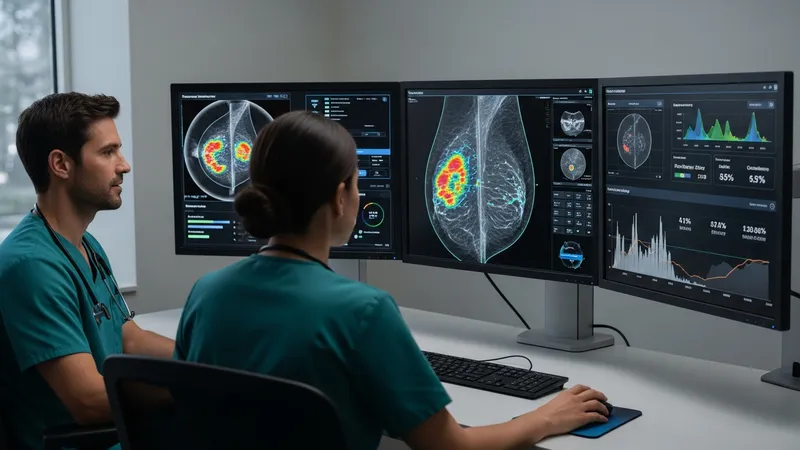
- Computer-aided detection for mammography and tomosynthesis: software that highlights suspicious regions on breast images to support radiologist review.
- AI-assisted digital pathology platforms: image-analysis tools that quantify histologic features and provide structured outputs for pathologists.
- EHR-integrated clinical decision support and predictive models: systems that combine clinical, imaging, and molecular data to generate risk scores or care-relevant summaries.
When comparing these example tool types, it is important to view them as complementary components rather than replacements for clinical expertise. Imaging detection software often focuses on sensitivity for certain lesion types but may vary by dataset and device. Digital pathology algorithms can standardize some quantitative measures but typically require human pathologist confirmation. EHR-integrated models that incorporate multiple data sources may improve situational awareness but are sensitive to the completeness and formatting of input data. Transparency about training data and intended use cases is a practical evaluation point.
Data quality and representativeness are central considerations for these computational methods. Models trained on limited or homogeneous datasets may perform differently when applied to diverse patient groups or imaging equipment. Validation studies often report performance on specific cohorts; readers should note that published results may reflect controlled datasets. Independent external validation and peer-reviewed evaluations are commonly cited practices to understand how a given method may behave in broader clinical settings.
Regulatory and implementation contexts influence how these systems are adopted in clinical practice. Depending on jurisdiction, some algorithms intended to inform diagnosis or treatment planning may require regulatory review or clearance. Operational integration with imaging systems, laboratory workflows, and electronic records typically involves technical and clinical governance steps. Institutions often pilot new tools, assess workflow impact, and define oversight mechanisms to ensure the output is used as intended and documented appropriately.
Ethical and practical considerations such as interpretability, data privacy, and clinician oversight are integral to responsible use. Interpretability features may help clinicians understand which inputs influenced an output, while privacy protections govern how patient data are stored and processed. Ongoing monitoring, periodic re-evaluation of model performance, and mechanisms for reporting unexpected behavior are often described in guidance documents on clinical AI deployment.
In summary, artificial intelligence approaches in breast cancer care encompass imaging analysis, pathology support, predictive analytics, and clinical decision support tools that may assist teams in diagnosis, planning, and monitoring. These methods typically augment information available to clinicians rather than dictate care, and their utility depends on data quality, validation, and appropriate oversight. The next sections examine practical components and considerations in more detail.